Long-term super microscopy
Nanographenes enable longer observation times
The 2014 Nobel Prize in Chemistry was awarded for the development of super-resolution fluorescence microscopy, including STED (stimulated emission depletion) microscopy. This method can be used to observe processes, e.g. in cells, with particularly high resolution. Researchers at the Max Planck Institute have now further developed this method by replacing conventional fluorophores with nanographenes. This means that processes with a longer duration can now also be observed, overcoming a previous limitation of STED microscopy.
The resolution of conventional microscopes is limited to around 200 nm, as the physicist Ernst Abbe described in the 19th century. However, interesting processes take place on a length scale below this limit, particularly in biological cells. STED microscopy overcomes this limit and achieves a resolution up to ten times better than conventional methods.
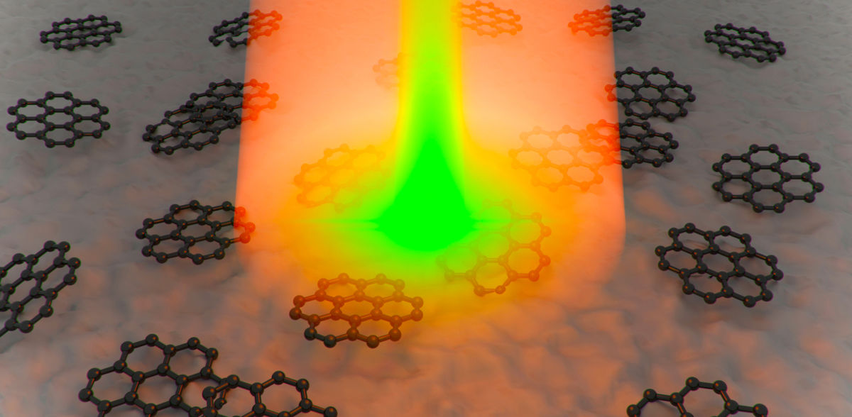
STED microscopy uses small fluorescent particles - fluorophores - in the sample, which glow (fluoresce) with the aid of an excitation laser. A second laser beam with a ring-shaped cross-section can deactivate the fluorescence in a ring-shaped area so that only a small central spot (less than 200 nm) remains illuminated. Scanning this beam combination across the sample produces a high-resolution image.
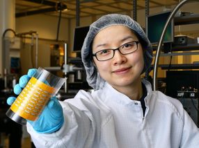
The main limitation of conventional STED microscopy has been the bleaching of fluorophores during prolonged illumination. This is particularly problematic when observing long-term processes that require repeated scanning. Researchers led by Xiaomin Liu at the MPI for Polymer Research, in collaboration with Akimitsu Narita and Ryota Kabe from the Okinawa Institute of Science and Technology, have solved this problem by using nanoscale nanographene particles. With nanographenes, the process of fluorescence fading can be reversed directly in the sample. For this purpose, the nanographene is illuminated with the ring-shaped beam: This illumination restores the nanographene's ability to fluoresce, so to speak.
This new method, which was presented in the renowned journal Nature Communications, opens up new possibilities for investigating previously unobservable processes using super-resolution microscopy. The ability to reactivate nanographenes with a high number of photons makes it ideal for long-term microscopy techniques and potentially expands its applications in biology and materials science.
Note: This article has been translated using a computer system without human intervention. LUMITOS offers these automatic translations to present a wider range of current news. Since this article has been translated with automatic translation, it is possible that it contains errors in vocabulary, syntax or grammar. The original article in German can be found here.
Original publication
Qiqi Yang, Antonio Virgilio Failla, Petri Turunen, Ana Mateos-Maroto, Meiyu Gai, Werner Zuschratter, Sophia Westendorf, Márton Gelléri, Qiang Chen, Goudappagouda, Hao Zhao, Xingfu Zhu, Svenja Morsbach, Marcus Scheele, Wei Yan, Katharina Landfester, Ryota Kabe, Mischa Bonn, Akimitsu Narita, Xiaomin Liu; "Reactivatable stimulated emission depletion microscopy using fluorescence-recoverable nanographene"; Nature Communications, Volume 16, 2025-2-4
Other news from the department science


These products might interest you
DM8000 M & DM12000 M by Leica
See More, Detect Faster
High-throughput Inspection Systems
LUMOS II by Bruker
FT-IR microscopy in the fast lane - the LUMOS II
One infrared microscope for all

alpha300 R by WITec
3D Raman microscopes with unequalled speed, sensitivity and resolution
Visualize and characterize every chemical detail
HYPERION II by Bruker
FT-IR and IR laser imaging (QCL) microscope for research and development
Analyze macroscopic samples with microscopic resolution (5 µm) in seconds

ZEISS ZEN core by Carl Zeiss
ZEISS ZEN core - Your Software suite for connected microscopy in laboratory and production
The comprehensive solution for imaging, segmentation, data storage and analysis