Minerals and ores in a chemical photo lab
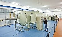
A unique colour X-ray camera goes into operation at the Helmholtz-Zentrum Dresden-Rossendorf (HZDR) today. With this camera, it will be possible for the researchers at the Helmholtz Institute Freiberg for Resource Technology (HIF), a part of the HZDR, to determine within a very short period of time the concentrations of such very finely dispersed metals as rare earth elements in ore minerals. Today, the scientists celebrate the start of the camera’s routine operation together with colleagues, partners, and companies who participated in the assembly of the camera. It was developed specifically to meet the institute’s analytical requirements.

Oliver Killig
“We want to use our colour X-ray camera primarily for the analysis of such trace elements as rare earth elements,” says the Head of the HIF’s Ion Beam Analytics Group, Dr. Axel Renno. These elements are dispersed very finely, and they usually occur only in small quantities. But they are very important for specific applications. This includes such elements as neodymium which is needed for the production of powerful magnets that are used in wind turbines; or cerium which is used in the form of cerium dioxide for catalytic converters, suntan lotions, or medications. Rare earths are one of the research focal points of the Helmholtz Institute Freiberg for Resource Technology; the researchers seek to extract raw materials from ore and recycle valuable resources from such discarded products as energy saving lamps. In addition to rare earth elements, there are also materials which are even scarcer, the so-called ultratrace elements. They play a vital role in explaining the processes associated with the formation of raw material deposits. The analysis of these elements will be another focus of the measurements taken by the colour X-ray camera.
Like any camera, the device has a detector chip; this is complemented by special optics for the X-rays. The camera analyzes with great precision both the chemical and the spatial composition of a sample. “The advantage is that we can now analyze materials much faster than with similar methods,” notes Axel Renno. For example, the analysis of a mineral sample can be reduced from one day to just one hour. This is made possible because the sample will not be “scanned” one sequence at a time, but will instead be fully illuminated and examined all at once.
First, a proton beam – i.e. fast, positively charged particles from the large-scale ion accelerator operated by the HZDR – is evenly directed onto a sample. When the protons hit the sample, X-rays are generated. They are characteristic for each and every element present in the sample and are recorded by the colour X-ray camera. This way, the researchers find out what chemical elements are in the sample. During this process, 70,000 pixels are simultaneously recorded in an image field that is 12 millimeters long. An optical image is created with the help of a special lens for X rays: Special capillary optics were developed from wafer-thin glass tubes; these optics assign each separate X-ray from the sample to the respective pixels.
The HZDR’s colour X-ray camera was subsidized by the Federal Ministry of Education and Research (BMBF) within the scope of the support measure “r3 – Strategic Metals and Minerals.” It is one of the few devices around the globe which permits the rapid analysis of the spatial distribution of such trace elements as rare earth elements. Unique, however, is the fact that the camera is operated with a proton beam generated by an ion accelerator, such as the one available at the HZDR. The camera was specifically adapted for these purposes. “We’d like to thank our colleagues at the Federal Institute for Materials Research and Testing (BAM) and the HZDR as well as at the participating companies: The Institute for Scientific Instruments GmbH, PN Sensor GmbH, DREEBIT GmbH, and TSO Thalheim Spezialoptik GmbH corporations, for their support and their expertise,” notes Renno.
So far, these types of cameras have only been operated at even larger accelerator systems, the so-called synchrotrons. Within the scope of the BMBF’s MEGA project, the Helmholtz Institute Freiberg (HIF) focuses on adapting the technology to the conditions prevalent in mines and processing plants. “We’re, thus, applying the advantages of ion beam analytics, just like we do here in Dresden, to the analysis of raw materials,” says Renno. Modern, spatially resolved analytical methods are an important basis for the development of more efficient raw material technologies such as those researched at the HIF.
Most read news
Topics
Organizations
Other news from the department science

Get the chemical industry in your inbox
By submitting this form you agree that LUMITOS AG will send you the newsletter(s) selected above by email. Your data will not be passed on to third parties. Your data will be stored and processed in accordance with our data protection regulations. LUMITOS may contact you by email for the purpose of advertising or market and opinion surveys. You can revoke your consent at any time without giving reasons to LUMITOS AG, Ernst-Augustin-Str. 2, 12489 Berlin, Germany or by e-mail at revoke@lumitos.com with effect for the future. In addition, each email contains a link to unsubscribe from the corresponding newsletter.


