A fast 4D look into materials and substances
A project is developing an AI-based software framework that can process terabyte-sized 4D tomography data on a desktop PC
Advertisement
To visualize processes at the micrometer scale, such as the discharge of a battery, scientists, so far, have required powerful computers that took days, if not months, to analyze large amounts of 4D tomography data. In the joint project “KI4D4E”, researchers are planning an open source framework that can be used for evaluating and visualizing 4D tomography data on a commercially available PC in a very short time. The Federal Ministry of Education and Research (BMBF) is supporting the project with about EUR 2.5 million. The project is coordinated by the Institute of Computer Architecture and Computer Engineering (ITI) at the University of Stuttgart.
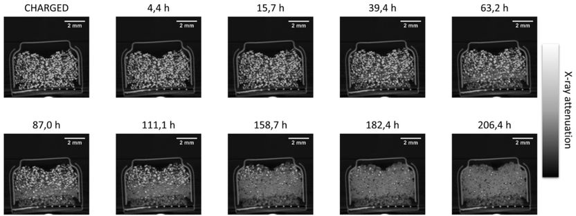
4D In-situ/Operando X-ray Tomography of a commercial Zn-Air Battery shows the dissolution of the zinc particles during disscharge.
Helmholtz-Zentrum Berlin (HZB), Tobias Arlt, Ingo Manke
How does the electrode of a lithium battery behave during discharge? How do bubbles grow in basaltic foam? How do bones change after inserting an implant? Researchers cannot answer questions like this by looking at a simple X-ray image. Instead, what they need are powerful synchrotron or neutron sources that allow them to obtain a temporal sequence of numerous 3D images (4D tomography). This quickly generates data volumes in the terabyte range, which, until now, researchers haven’t been able to analyze systematically, and only with a lot of manual work.
This is where the joint project “KI4D4E” (“An AI-based framework for the visualization and evaluation of massive amounts of 4D tomography data for beamline end users”) comes in. The partners involved are planning a framework that is already running on commercially available PCs for gaming applications and with 128 Gbyte RAM. The terabyte-sized data volumes are to be compressed and stored in RAM, and then processed and visualized in real time. The researchers want to correct artifacts that occur during tomography – such as noise or motion blur – with novel artifact reduction algorithms. Artificial intelligence (AI) with deep neural networks will be used for this purpose.
Subsequent time- and location-independent evaluation
In the future, end users will gather data on their materials, substances, and objects as usual, that is at a stationary neutron source or beamline of a powerful large-scale research device (synchrotron beamlines). Once the measurement data has been collected, however, they will be able to evaluate it subsequently and independently of time and location, requiring just a suitably equipped desktop PC at their own institute.
The basis for the planned software framework is an application program that is available as open source and can be extended to display images of three-dimensional objects. This so-called 3D voxel viewer was developed at Faculty 5 (Computer Science, Electrical Engineering and Information Technology) of the University of Stuttgart in 2014. There, the joint project is coordinated by the Department of Computational Imaging Systems at the Institute of Computer Architecture and Computer Engineering (ITI). Project partners include the Karlsruhe Institute of Technology, the Fraunhofer Society for the Promotion of Applied Research, the Forschungszentrum Jülich national research institution, the University of Erlangen-Nuremberg, the Helmholtz Center for Energy and Materials Research in Berlin, the Helmholtz Center Hereon, and the University of Passau.


