With light into the nanoworld: How optical microscopes allow detailed investigations of nanoparticles
The goal: the development of novel nanoparticles for biomedical applications.
It sounds like trying to scan a vinyl record with a hammer: light is actually too "coarse" to image small particles on the nanometer scale. However, in their project "Supercol"- funded by the European Union - scientists want to achieve just that: The investigation of nanoparticles with light. To make this possible, they are combining Nobel Prize-winning methods with modern computer processes. The goal: the development of novel nanoparticles for biomedical applications.
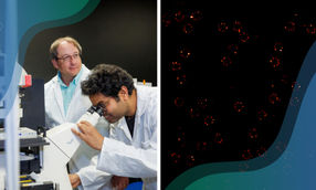
Using a combination of super-resolution microscopy and electron microscopy, scientists can now determine the position of molecules on the surface of nanoparticles much more precisely. In the future, this could enable new biomedical applications.
Max-Planck-Institut für Polymerforschung
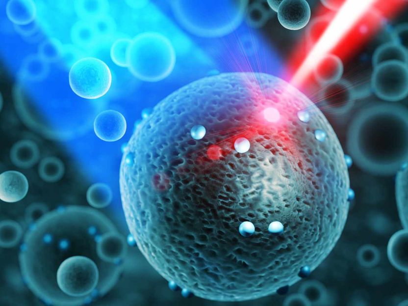
Nanoparticles - i.e. small particles with a size in the range of a few tens to hundreds of billionths of a meter - are a wide-ranging field of research. For example, they make the latest biomedical applications possible by acting as a kind of container to transport active ingredients to their target. Ideally, their surfaces are "functionalized" - i.e. provided with a molecular puzzle piece, which allows them to dock only to desired target cells in the body.
However, studying such particles and the molecules on their surface is difficult: light is basically too "coarse" to image such particles in a normal light microscope. Visible light in the range from UV to infrared can at most resolve particles with a size of 200 nanometers - 200 billionths of a meter. Too large to determine where, for example, a molecular puzzle piece sits on its surface or to determine their number.
"It's kind of like trying to listen to a record with a hammer, when what you really need is a needle," explains Ingo Lieberwirth, group leader in Katharina Landfester's department at the Max-Planck-Institute for Polymer Research. Lieberwirth leads the "Electron Microscopy" group and therefore knows that "electron microscopes can image such particles well - but there is also a great risk that the electrons used will damage the docked molecules."
On to higher resolution
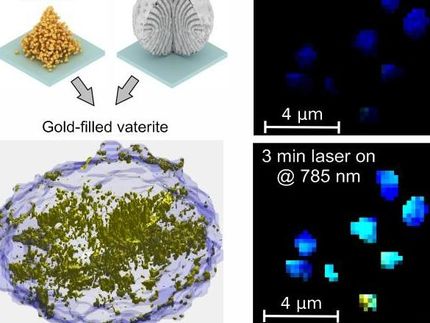
Therefore, the researchers used a method that won them the 2014 Nobel Prize in Chemistry: In what is known as "super-resolution microscopy," small fluorescent particles - called fluorophores - are used and, in the case of nanoparticles, attached to molecules on its surface. These fluorophores have the property of blinking statistically in a microscope. The position of this blinking signal can be detected much more accurately than would be possible with conventional optical microscopy.
"Just think of it like two people standing side by side on a dark mountain, shining their flashlights in their direction," Lieberwirth said. "If they're both shining at the same time, it's hard to tell, that there are two flashlights. But when it's blinking, the difference in position becomes much clearer."
Using computer power to get to the truth
However, the image of the nanoparticle obtained in this way is only half the truth: Nanoparticles have properties that can distort this image - for example resonance phenomena that bring also part of the nanoparticle to glow, and not only the fluorophore. The scientists therefore imaged nanoparticles using both electron microscopy and super-resolution optical microscopy. While electron microscopy provides the "true" position of the docked molecule, physical effects in the light microscope lead to a shift. Software now correlates both images - and can thus predict the true position based on the light microscope image.
The researchers now hope to use their method to study nanoparticles in the light microscope, which delivers faster results and does not destroy the particles. This will allow nanoparticles to be studied more precisely and comprehensively in the future, leading to new biomedical applications.

Other news from the department science


These products might interest you
NANOPHOX CS by Sympatec
Particle size analysis in the nano range: Analyzing high concentrations with ease
Reliable results without time-consuming sample preparation

Eclipse by Wyatt Technology
FFF-MALS system for separation and characterization of macromolecules and nanoparticles
The latest and most innovative FFF system designed for highest usability, robustness and data quality

DynaPro Plate Reader III by Wyatt Technology
Screening of biopharmaceuticals and proteins with high-throughput dynamic light scattering (DLS)
Efficiently characterize your sample quality and stability from lead discovery to quality control