CU physicists use ultra-fast lasers to open doors to new technologies unheard of just years ago
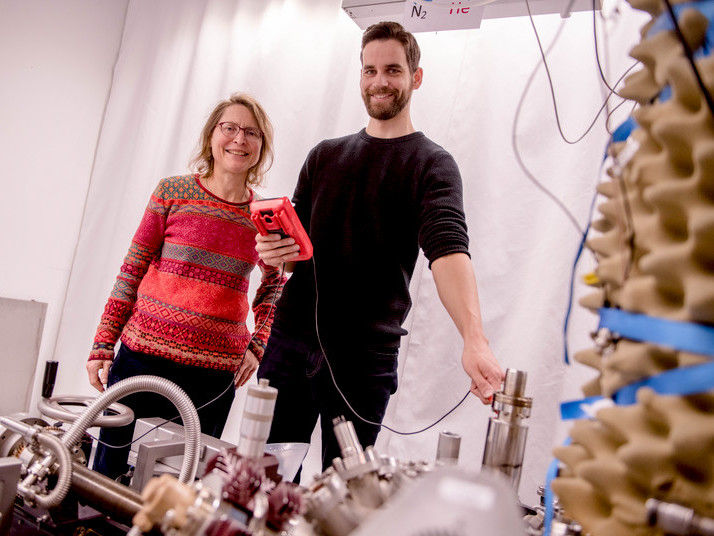
For nearly half a century, scientists have been trying to figure out how to build a cost-effective and reasonably sized X-ray laser that could, among other things, provide super high-resolution imaging. And for the past two decades, University of Colorado at Boulder physics professors Margaret Murnane and Henry Kapteyn have been inching closer to that goal.
Recent breakthroughs by their team at JILA, a joint institute of CU-Boulder and the National Institute of Standards and Technology, have paved the way on how to build a tabletop X-ray laser that could be used for super high-resolution imaging, while also giving scientists a new way to peer into a single cell and gain a better understanding of the nanoworld. Both of these feats could lead to major breakthroughs in many fields including medicine, biology and nanotechnology development.
"Our goal is to create a laser beam that contains a broad range of X-ray wavelengths all at once that can be focused both in time and space," Murnane said. "If we have this source of coherent light that spans a huge region of the electromagnetic spectrum, we would be able to make the highest resolution light-based tabletop microscope in existence that could capture images in 3-D and tell us exactly what we are looking at. We're very close."
Murnane and Kapteyn presented highlights of their research at the American Association for the Advancement of Science, or AAAS, annual meeting in San Diego, during a panel discussion about the history and future of laser technology titled "Next Generation of Extreme Optical Tools and Applications."
Most of today's X-ray lasers require so much power that they rely on fusion laser facilities the size of football stadiums or larger, making their use impractical. Murnane and Kapteyn generate coherent laser-like X-ray beams by using an intense femtosecond laser and combining hundreds or thousands of visible photons together. And the key is they are doing it with a desktop-size system.
They can already generate laser-like X-ray beams in the soft X-ray region and believe they have discovered how to extend the process all the way into the hard X-ray region of the electromagnetic spectrum.
"If we can do this, it could lead to all kinds of possibilities," Kapteyn said. "It might make it possible to improve X-ray imaging resolution at your doctor's office by a thousand times. The X-rays we get in the hospital now are limited. For example, they can't detect really small cancers because the X-ray source in your doctor's office is more like a light bulb, not a laser. If you had a bright, focused laser-like X-ray beam, you could image with far higher resolution."
Their method can be thought of as a coherent version of the X-ray tube, according to Murnane. In an X-ray tube, an electron is boiled off a filament, then it is accelerated in an electric field before hitting a solid target, where the kinetic energy of the electron is converted into incoherent X-rays. These incoherent X-rays are like the incoherent light from a light bulb or flashlight -- they aren't very focused.
In the tabletop setup, instead of boiling an electron from a filament, they pluck part of the quantum wave function of an electron from an atom using a very intense laser pulse. The electron is then accelerated and slammed back into the ion, releasing its energy as an X-ray photon. Since the laser field controls the motion of the electron, the X-rays emitted can retain the coherence properties of a laser, Murnane said.l
Most read news
Organizations
Other news from the department science

Get the chemical industry in your inbox
By submitting this form you agree that LUMITOS AG will send you the newsletter(s) selected above by email. Your data will not be passed on to third parties. Your data will be stored and processed in accordance with our data protection regulations. LUMITOS may contact you by email for the purpose of advertising or market and opinion surveys. You can revoke your consent at any time without giving reasons to LUMITOS AG, Ernst-Augustin-Str. 2, 12489 Berlin, Germany or by e-mail at revoke@lumitos.com with effect for the future. In addition, each email contains a link to unsubscribe from the corresponding newsletter.
Most read news
More news from our other portals
Last viewed contents
Haplogroup_I1a_(Y-DNA)
WACKER Expands Technical Center in Singapore and Includes New Lab for Silicone Elastomers
Next generation of high clarity copolyester introduced by Eastman
Heisenberg_model_(classical)
Mechanically imprinting atoms in ceramic
