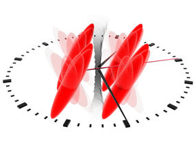
The first atomic X-ray laser
Advertisement
A group of scientists headed by Nina Rohringer from the Hamburg Center for Free-Electron Laser Science (CFEL) realized the first X-ray laser based on atoms at the Californian research centre SLAC. Using neon atoms, they generated ultra-short X-ray bursts of unique colour purity. In many cases, this allows a sharper look into the nano world, as scientists reported in the British journal “Nature” (DOI: 10.1038/nature.10721). CFEL is a joint enterprise of Deutsches Elektronen-Synchrotron DESY, the Max Planck Society and the University of Hamburg.
Max Planck scientist Rohringer and her colleagues from Lawrence Livermore National Laboratory and Colorado State University used the so-called free-electron laser LCLS at SLAC for their experiments. With strong magnets, free-electron lasers bring fast electrons accelerated by particle accelerators on a zigzag course, thus generating laser like radiation within the X-ray range. Unlike these, traditional optical lasers are based on the radiation of atoms which are excited to emit light by simulated emission. . So far, this was not possible in the X-ray range, because the excitation of atoms in this area requires very intensive radiation. With the LCLS, Rohringer and her team now created the first X-ray laser based on atoms – more than 40 years after the original concept was published.
Due to their short wave lengths, X-ray lasers are able to make atomic details visible and, with their ultra-short pulse duration, take snapshots of fast molecular processes. Thus, it may be possible to photograph the process of chemical reactions. The purer the colour of the laser and the shorter the X-ray burst, the sharper will be the image.
The scientists sent a short LCLS X-ray pulse of 40 to 80 femtoseconds (one femtosecond is one quadrillionth of a second) through a neon gas cell at high pressure. The X-ray beam cut a narrow channel through the gas, along which neon atoms were ionised. In this process, an inner-shell electron was kicked out of the atom, leaving a hole behind. Subsequently, one of the electrons of the outer shell filled up the hole in the inner shell, thereby emitting an X-ray pulse. According to the self-amplifying effect, this pulse stimulated the next atom to emit an X-ray pulse – an avalanche effect – so that the numerous pulses overlap and form one X-ray laser burst. The wavelength of this X-ray light was 1.46 nanometres (millionth of a millimetre). For comparison: most applied lasers in the optical range have a wavelength of 800 nanometres. The wavelength determines the size of the details which are still discernible in the corresponding light.
“The generated X-ray light is a bit weaker than that of the free-electron laser, but it has a more stable wavelength, a smoother pulse profile and a shorter pulse length,” Rohringer explains. Also free-electron lasers have a sharply defined colour. The energy – or the wavelength – of its X-ray radiation fluctuates within the range of about 15 electronvolts, at an energy of about 1000 electronvolts. The energy width of the X-ray flash of neon atoms was only 0.25 electronvolts – this is 60 times sharper.
The X-ray flashes from the free-electron laser und from neon atoms have different wavelengths. This creates a two-colour X-ray laser, with an optimal synchronisation of both pulses. This, for example, may be used to start a process with one pulse - e.g. a chemical reaction or an excitation or a structural change in a solid state – and then take a photograph of this process with the pulse of the other colour after a certain time. When the pulse is directed via a precisely defined detour, it is possible to delay it for a required short period of time, in order to photograph different stages of a chemical reaction. Since both pulses are generated at the same time, this period of time can be defined with high accuracy.
At CFEL in Hamburg, Rohringer currently investigates how this technique may be expanded. “We are exploring for example how to reach higher energies, and whether it is possible to use molecules, for example oxygen, instead of neon atoms as a laser medium.”
Original publication
“Atomic inner-shell x-ray laser at 1.46nm pumped by an x-ray free electron laser”; Nina Rohringer, Duncan Ryan, Richard A. London, Michael Purvis, Felicie Albert, James Dunn, John D. Bozek, Christoph Bostedt, Alexander Graf, Randal Hill, Stefan P. Hau-Riege and Jorge J. Rocca; Nature, Bd. 481, S.488


